Fast, accurate tissue sample analysis speeds up the work of pathologists and researchers and ensures better patient care.
Analysing tissue samples the traditional way – slowly and strenuously, while hunched over a microscope – may now be a thing of the past. Pathologists and researchers can accelerate and automate the analysis process using tools developed by Fimmic, a Finnish startup founded in 2013: Aiforia is a virtual microscope and a cloud platform in the same software. Fimmic is a spin-off of the Finnish Institute for Molecular Medicine at the University of Helsinki.
“Our deep-learning AI image analysis technology enables fast and accurate automation of complex image analysis tasks not previously possible,” says CEO Kaisa Helminen.
“Our AI software is trained to detect and quantify objects, categorise cancer tumours based on progression, and identify rare targets such as malaria parasites,” she Helminen. “For the first time, we’re able to mimic a human observer in understanding the context in tissue.
“The solution acts as a tireless analysis support tool, or like a second opinion, for pathologists and researchers, speeding up the workflow and preventing human errors in interpretation. This way, it ensures better patient care.”
Results in minutes

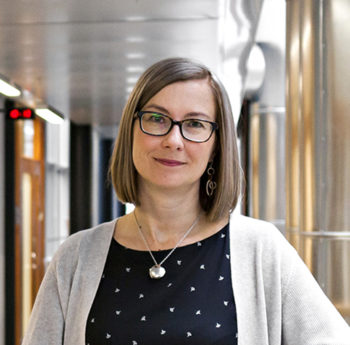
“For the first time, we’re able to mimic a human observer in understanding the context in tissue,” says Kaisa Helminen of Fimmic.Photo: Sebastian Mardones/Health Capital Helsinki
The on-demand process runs in a cloud computing environment. The platform operates on a software-as-a-service basis, meaning customers do not need to buy local hardware or install any local software. All they have to do is upload their scanned tissue sample images to the service, and the results will arrive in minutes.
“In 2018, Aiforia will be used for analysing clinical patient samples for the first time,” Helminen says. “There is also a big need for this type of software in the early preclinical phase of new drug development.”
Investors agree; the company closed a five-million-euro funding round in November 2017.
By Leena Koskenlaakso, ThisisFINLAND Magazine 2018


